
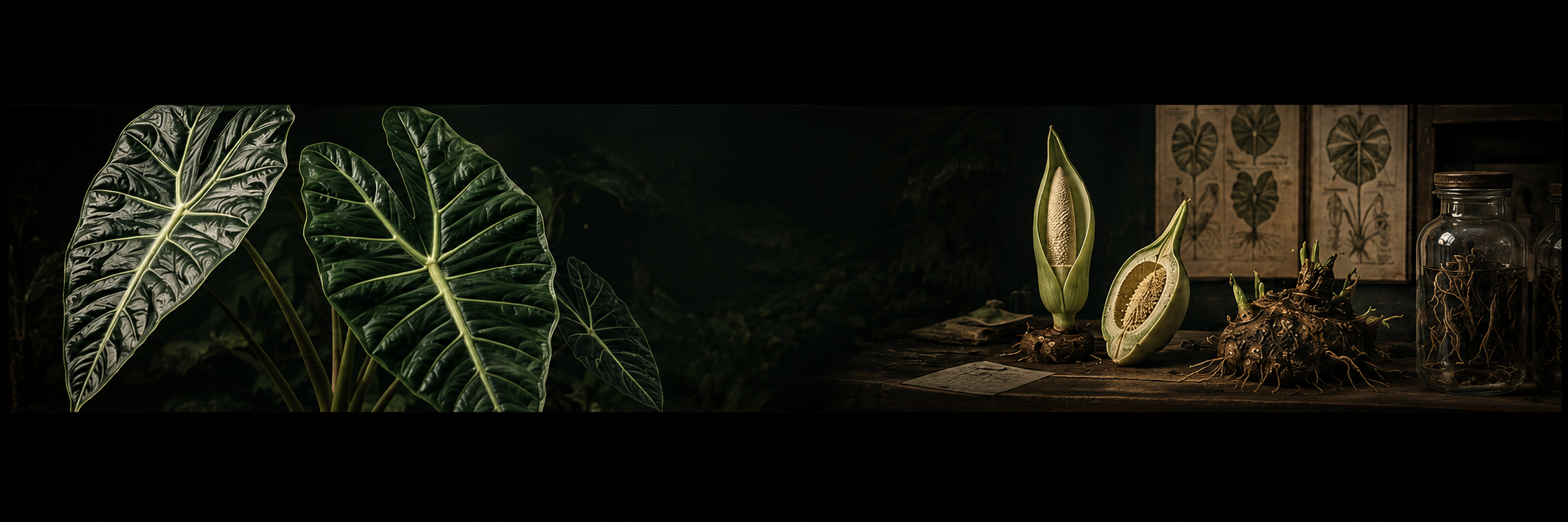
Araceae · Morphology
THE
ALOCASIA LEAF
How to read shape, venation and character
Opening
A leaf is not a name.
A leaf is evidence.
At first glance, a leaf may seem simple: a shape, a color, and perhaps a conspicuous pattern of venation. In Alocasia, a leaf carries considerably more information than that.
Taxonomic descriptions do not stop at the overall outline. They consider the division of the blade, the development of the anterior and posterior lobes, the costae, the several distinct categories of veins, and — above all — the course those veins follow and the connections they make within the blade. In species that look alike at first sight, these characters are often where the real differences live.
A leaf can tell us a great deal. But not always everything.
Which side are you looking at?
Before a leaf can be described, it must be clear which surface is being observed. The adaxial surface is the upper surface of the blade — the side that faces the shoot axis as the leaf develops. The abaxial surface is the lower surface, the side that faces away.
This distinction matters more than it may appear. A vein can be impressed on the upper surface and distinctly raised on the lower surface of the same leaf. Color, glands and surface texture routinely differ between the two sides — in Alocasia the glands in the vein axils sit on the abaxial surface1, and many species carry their most striking coloration there. When a botanical description states that a character is adaxial or abaxial, that word is part of the character itself.
Turn the leaf over
And rather than beginning with “what plant does this leaf look like?”, begin with the question a taxonomist asks: what morphological characters does this leaf actually possess?
Part I
The map of the blade
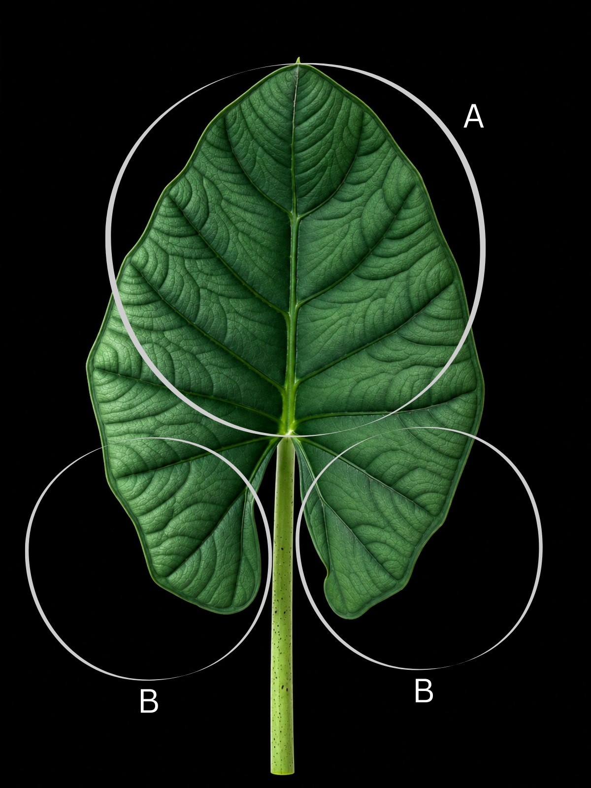
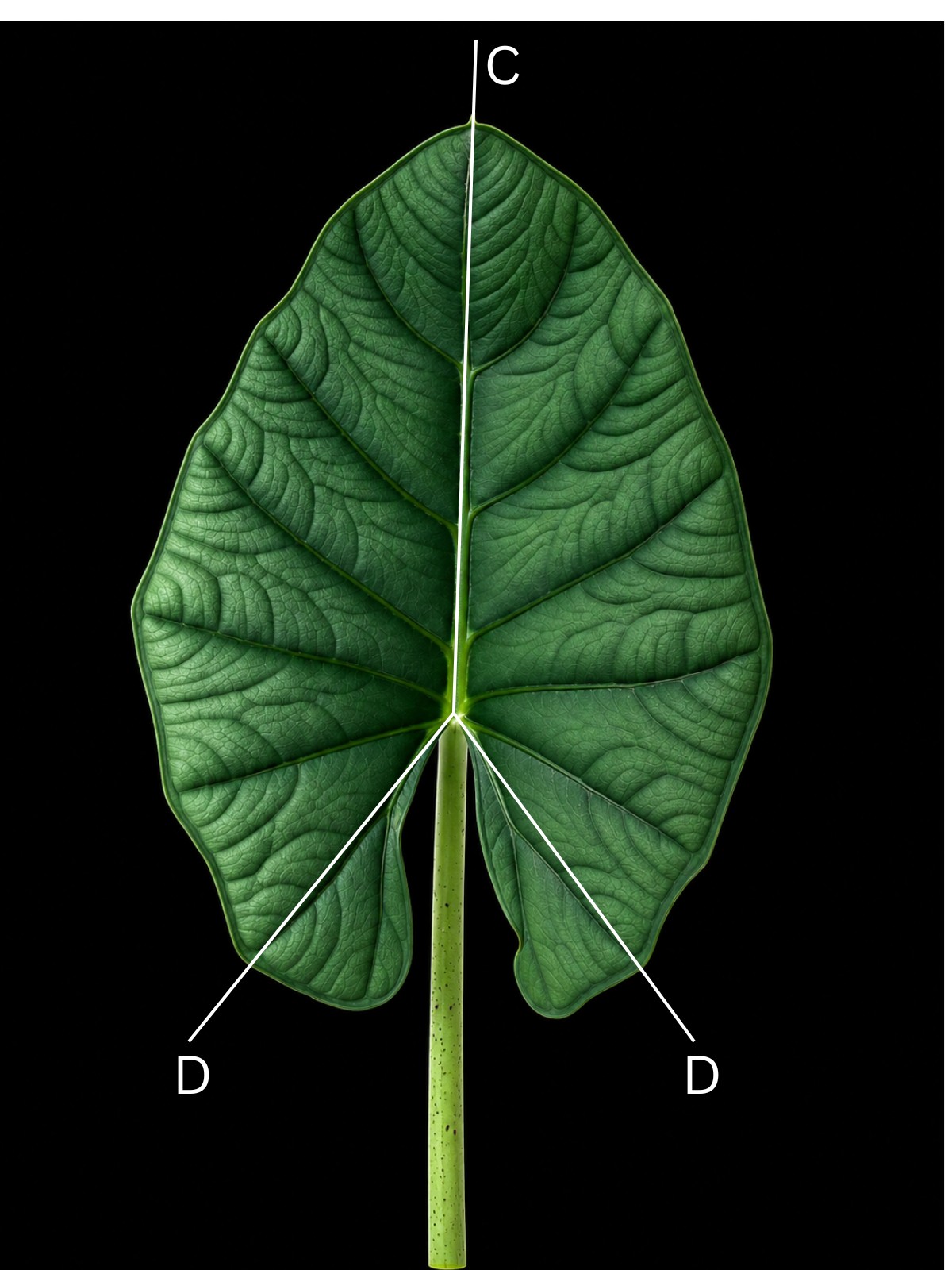
For describing the Alocasia leaf, the revision of the genus for West Malesia and Sulawesi by Alistair Hay1 provides the standard reference diagram, and its lettering scheme is the vocabulary used throughout this article. The blade is not treated as a heart-, shield- or arrow-shaped surface. It is a set of distinguishable regions carrying an organized system of venation.
Photographs by Nadine Hafke
A — anterior lobe. The large forward portion of the blade, extending toward the apex. It usually forms the greater part of the lamina.
B — posterior lobes. The two basal lobes. Their size, shape, orientation and proportions vary considerably among species — which is exactly why taxonomic descriptions measure them rather than merely naming the overall outline.
C — anterior costa. The strong central rib of the anterior lobe, running toward the apex.
D — posterior costae. The main axes of the two posterior lobes, one running through each lobe. They are not simply extra lateral veins: each is the structural midrib of its lobe, and the primary veins of the posterior lobe run pedately off its outer side1 — a different arrangement from the pinnate venation of the anterior lobe. The angle at which the posterior costae diverge, and whether they run straight or curve, are characters that appear again and again in species descriptions.
The rest of the scheme
The remaining letters cover the vein categories, the glands, the margin and the base of the leaf. Each is taken up in its own section below.
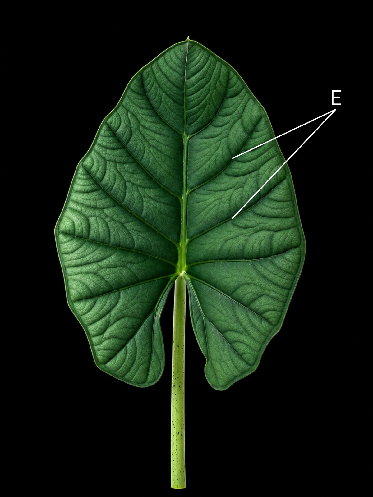
- E. Primary lateral vein.
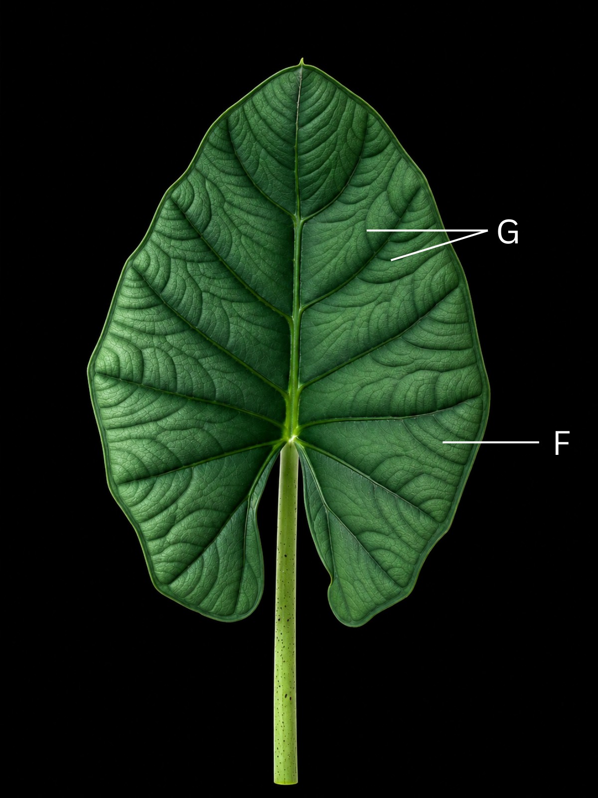
- F. Subsidiary vein (with axillary gland).
- G. Secondary vein.
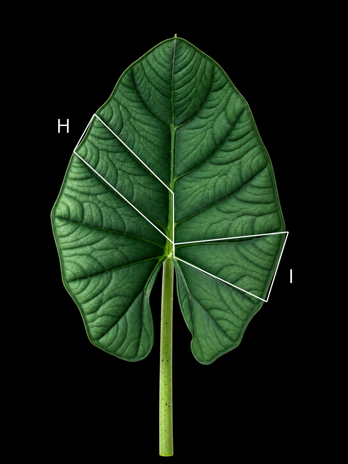
- H. Sector with interprimary collective vein formed by meeting of secondary veins.
- I. Sector with interprimary collective vein not formed.
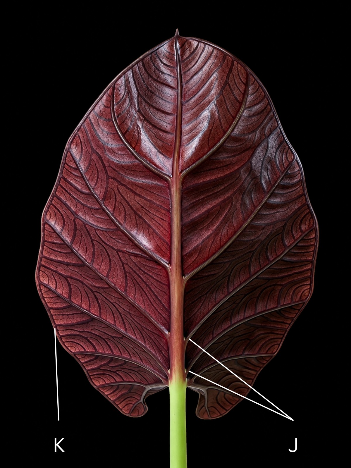
- J. Axillary gland.
- K. Intramarginal vein.
- L. Inner side of posterior lobe.
- M. Petiole.
- N. Sinus.
The proportions of these regions are themselves characters: length and width of the anterior lobe, length and shape of the posterior lobes, their orientation, the ratio of anterior to posterior blade, and the shape and depth of the sinus. This is why “heart-shaped” or “arrow-shaped” alone is rarely enough for taxonomic comparison.
Part II
The venation hierarchy
Primary lateral veins (E)
The strong primary lateral veins are the most conspicuous element of the venation, running pinnately off both sides of the anterior costa1. When comparing leaves, descriptions consider their number, spacing, angle of divergence, course, relationship to the anterior costa, and behavior as they approach the margin.
Their number alone, however, is not a taxonomic fingerprint. Two species can share a count and differ in everything the count does not capture.
Subsidiary and secondary veins (F, G)
Between the primary lateral veins lies a second system, and Hay's scheme1 splits it in two. Subsidiary veins follow the general course of the secondary venation but are distinctly stronger than ordinary secondary veins — intermediate in thickness between the primary and secondary venation2. Not every conspicuous vein between two primaries is simply an “intermediate vein”; whether a species produces subsidiary veins at all can help characterize it. Secondary veins form the finer venation within the areas bounded by the stronger veins.
Photographs by Nadine Hafke
The secondary venation of Alocasia follows the pattern that aroid taxonomists call colocasioid venation, a signature of the tribes Colocasieae and Caladieae across the whole family: the finer veins branch from the primary lateral veins almost at right angles, arch strongly toward the leaf margin, often fuse along the way into a more-or-less sinuose interprimary collective vein, and finally join a submarginal collective vein8. In Hay's shorter formulation for Alocasia, the secondary veins leave the primaries at a wide angle and are then deflected toward the margin1.
This shared architecture is precisely why the interesting questions about any individual vein are not “is it there?” but: where does it originate, how strong is it, what course does it follow, and which other veins does it connect with?
Interprimary collective veins (H, I)
Of particular interest is what the secondary veins do between two neighboring primaries. In some sectors of the blade they meet one another and unite into an interprimary collective vein. In other sectors no such vein forms, and the secondaries run separately to the margin. Hay's diagram deliberately illustrates both situations side by side1.
Photographs by Nadine Hafke
Whether interprimary collective veins form, and whether they run smoothly, undulate or zig-zag, are characters that separate real species — as the case studies below will show.
A character can be real and still invisible
In the Bornean species Alocasia chaii, the interprimary collective veins are well defined when the leaf is fresh but decidedly obscure in dried material9. A character can be plain on the living plant and nearly absent from a herbarium sheet — one more reason to record venation while the leaf is in front of you.
Venation is not a collection of visible lines. It has an architecture. How do the veins connect?
Part III
The margin and the glands
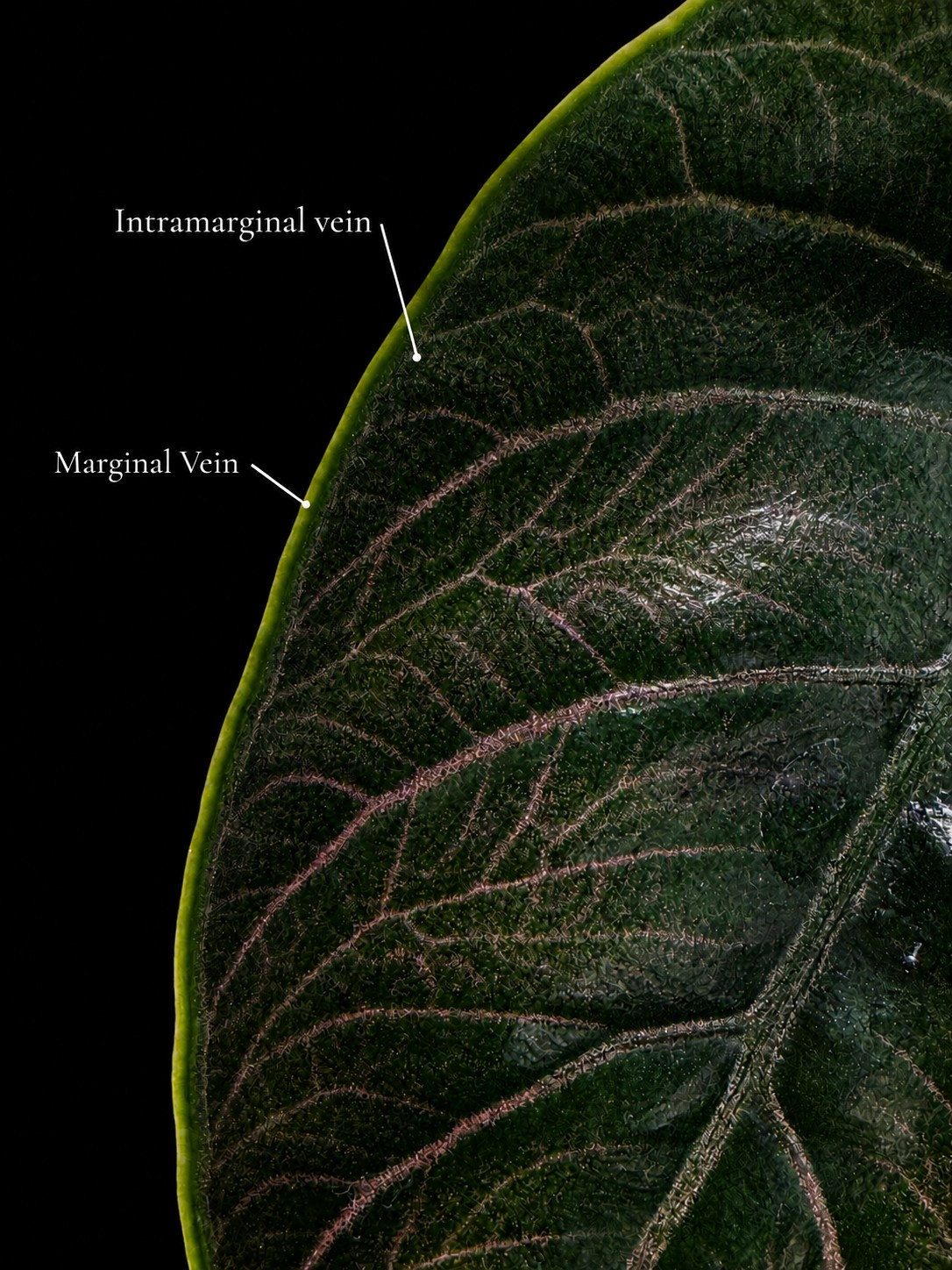
Intramarginal vein (K)
The intramarginal vein runs within the blade, close to the margin. It is not the leaf margin itself, and it should not automatically be equated with a marginal vein running at the very edge — some species carry both, a marginal vein at the edge and an intramarginal vein a few millimeters inside it3. In other species the major venation runs instead to a submarginal vein a short distance from the edge4. At genus level the Flora of China puts it concisely: the primary lateral veins form a submarginal collective vein, with one or two closely adjacent marginal veins also present10. What matters, whichever terms a description uses, is being able to follow each as an actual vascular structure within the lamina.
Axillary glands (J)
In the axils of the primary veins on the abaxial surface, Alocasia typically carries small glands1. The Australasian revision by Hay & Wise describes them as wax-secreting, and notes that they may also occur in the axils of secondary veins and scattered on the petiole5; the West Malesian revision is more reserved and calls their function unknown1. They can be conspicuous: in Alocasia suhirmaniana the glands in the primary-vein axils are a prominent purple2.
Photographs by Nadine Hafke
Their development also varies — between species and, in some cases, within one. For the New Guinea species Alocasia nicolsonii, Hay & Wise tabulate specimens whose abaxial wax glands range from plentiful to sparse to none5.
And like the veins, the glands can carry a diagnosis. In Boyce's key to the Alocasia of Borneo, Alocasia chaii is separated partly by conspicuous deep red axillary glands, Alocasia beccarii partly by inconspicuous pale green ones9. In the newest addition to the Sulawesi flora, Alocasia crispa is distinguished from Alocasia celebica partly by its conspicuous creamy axillary glands — and from Alocasia suhirmaniana by the same character set, along with the crisped, translucent leaf margin that gives the species its name11.
Not dots to be found in a photograph
Axillary glands are not simply small dots to be “found.” Their position, their actual development on the plant in hand, and the taxonomic context in which they are described are what carry the information.
Part IV · Case study
When a vein becomes diagnostic
The intramarginal vein is not merely a term in a diagram. In the right comparison, it is the diagnosis.
Alocasia azlanii was described from Brunei by Wong & Boyce in 2016 and compared explicitly with Alocasia beccarii and Alocasia peltata3 — all three of them small herbs with peltate, glabrous leaves, part of the same alliance of diminutive species as the horticulturally famous Alocasia cuprea3. The description follows Hay's terminology1, and its distinguishing characters map directly onto the diagram letters above.
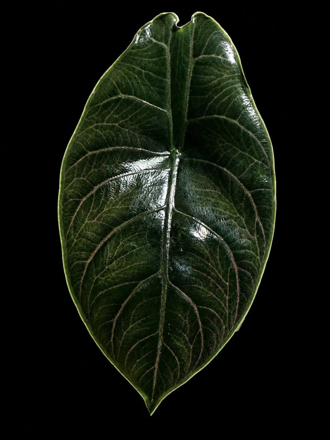
In Alocasia azlanii, both a conspicuous marginal vein and an intramarginal vein are developed. The intramarginal vein runs about 2–4 mm inside the margin, inward of the marginal vein. The primary lateral veins of the anterior lobe — 2 or 3 on each side of the anterior costa — diverge at about 45–60° and curve upward into the intramarginal vein. The secondary veins are confluent: they meet and form interprimary collective veins3.
Photographs by Nadine Hafke
The living leaf demonstrates directly that the intramarginal vein is not the outermost edge of the blade but a vascular structure running within the lamina.
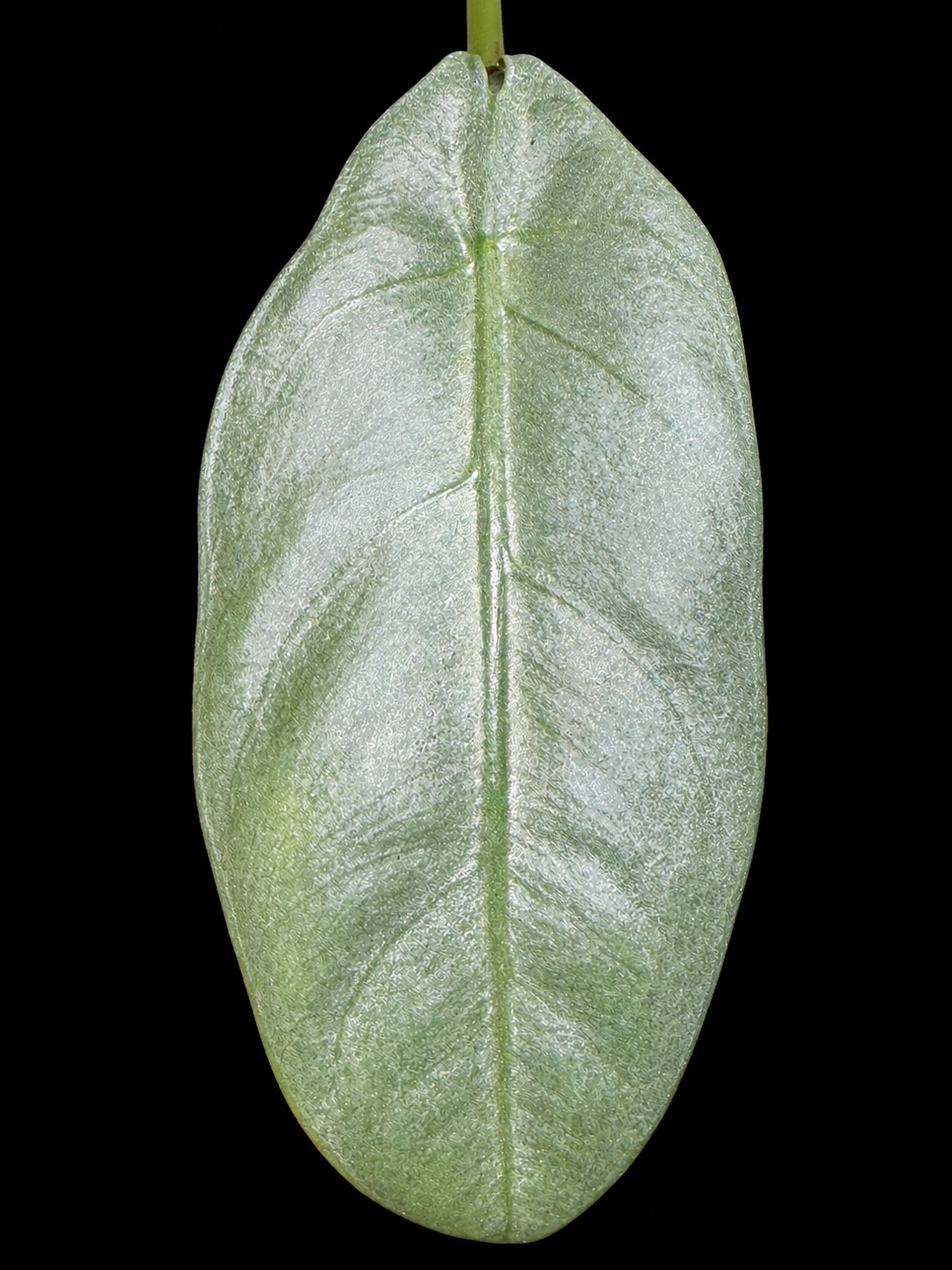
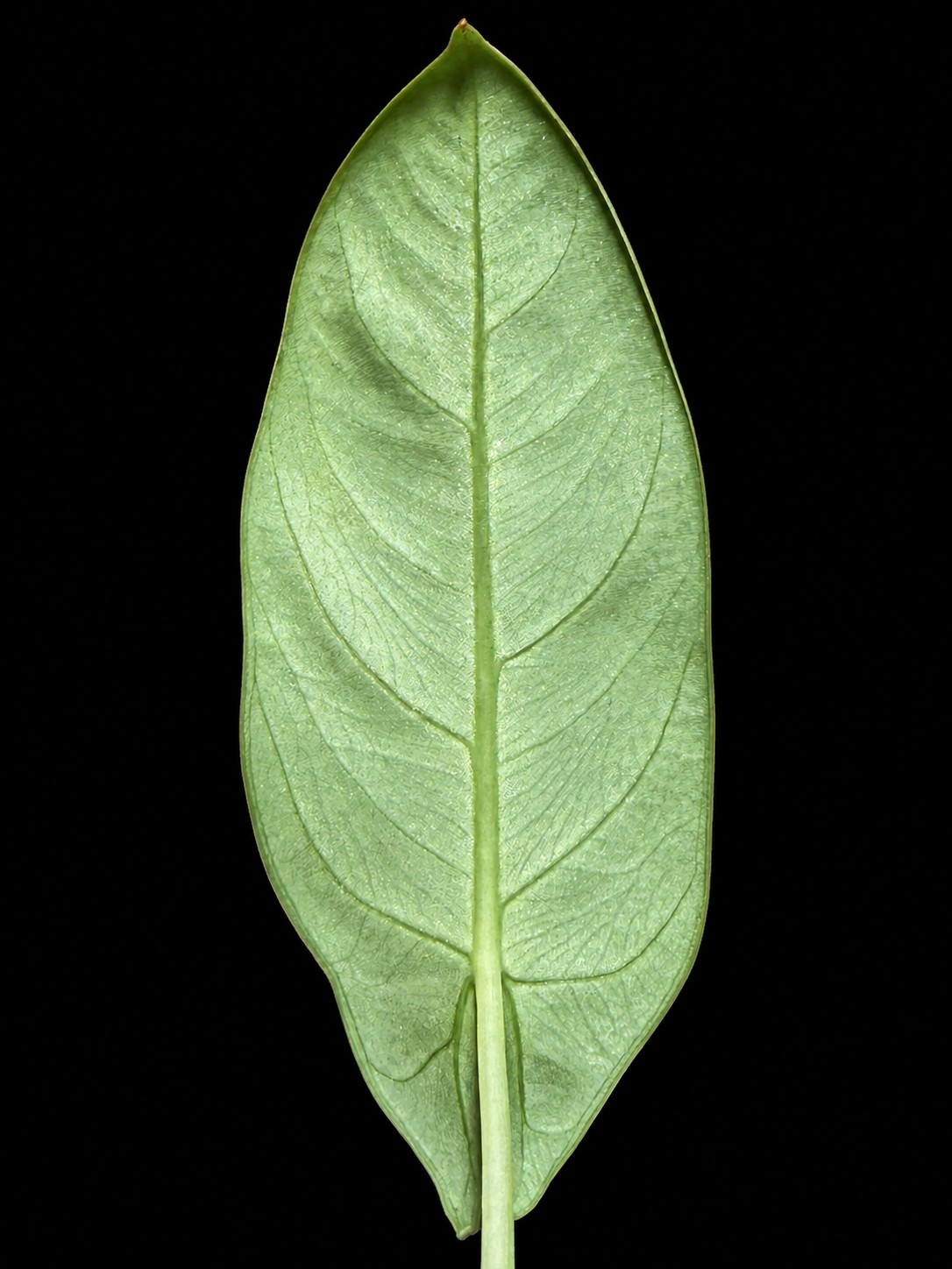
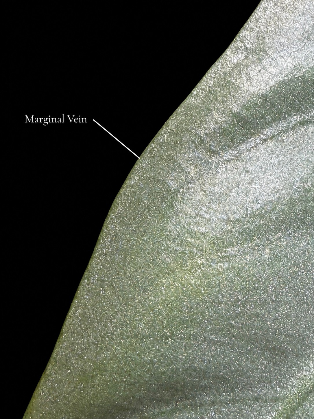
In Alocasia beccarii, the venation near the margin is organized differently. Wong & Boyce separate Alocasia azlanii from Alocasia beccarii precisely by those conspicuous marginal and intramarginal veins — in Alocasia beccarii only a clear marginal vein is evident3.
That wording deserves attention. It does not claim that the total absence of every possible intramarginal vascular connection can be proven from a photograph. What matters is the diagnostically different expression of the visible marginal venation that Wong & Boyce used.
Photographs by Nadine Hafke
Photographs by Nadine Hafke
Alocasia beccarii carries its own quiet leaf characters elsewhere: in the Bornean key it is recognized partly by inconspicuous pale green axillary glands9 — the same structures from the previous section, doing diagnostic work in another comparison.
And a second lesson in the same paper
Against Alocasia peltata, Wong & Boyce use the behavior of the secondary veins: in Alocasia azlanii they are confluent and form interprimary collective veins; in Alocasia peltata they do not meet in this way, and interprimary collective veins are not formed3.
That is exactly the H-versus-I contrast of Hay's diagram — one species is the H sector, the other is the I sector, and the difference is diagnostic.
The principle
2–4 mm
A seemingly minor feature of leaf venation — a vein a few millimeters inside the edge — can form part of the formal distinction between similar species. Don't just name the vein. Ask what it tells you.
Part V
Sinus, lobes and petiole
N — the sinus is the indentation or space between the two posterior lobes. It is not the point from which the major veins radiate — that point is the petiole insertion.
L — the inner side of posterior lobe is the side of a posterior lobe facing the sinus. The distinction exists because a posterior lobe is not symmetrical about its posterior costa, so descriptions often characterize the inner piece of lamina separately — “inner side of posterior lobe lanceolate” describes only that inner strip1.
M — the petiole is the leaf stalk, connecting the blade to the rest of the shoot.
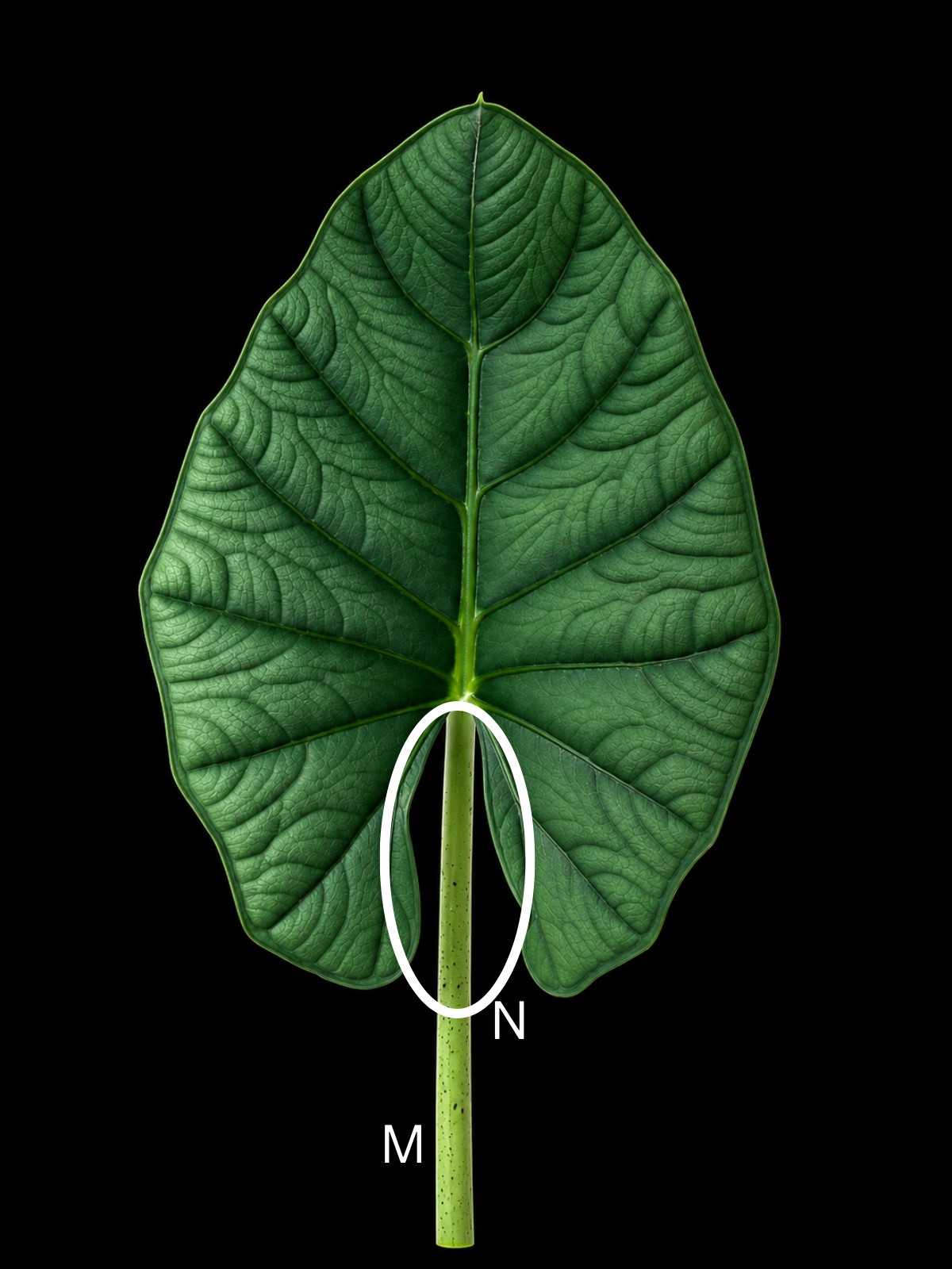
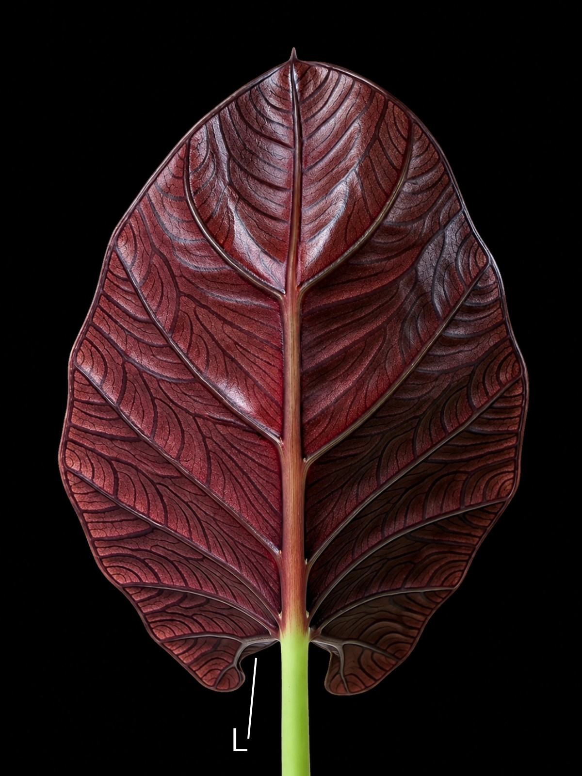
Photographs by Nadine Hafke
In peltate leaves the picture changes: the posterior lobes are joined across the sinus, sometimes almost completely — in Alocasia azlanii they are united except for a 4–8 mm incision at the extreme base of the blade3. A description will then say how far the lobes are joined, and whether the posterior costae are “naked” — running without lamina — for part of their length in the sinus4.
From diagram to description: Alocasia suhirmaniana
The original description of Alocasia suhirmaniana from Sulawesi by Yuzammi & Hay shows just how much of a species description is written in the diagram's vocabulary2.
The blade is broadly ovato-sagittate and peltate, pointed downward, glossy dark green above and dark purple beneath. The anterior lobe reaches about 35 cm. Up to eight primary lateral veins run on each side of the anterior costa — the proximal ones diverging at 70–80°, the distal ones at about 45° — with conspicuous purple glands in their axils on the abaxial side2.
Each primary vein of the anterior lobe carries 1–3 subsidiary veins, thicker than the secondary venation whose course they follow. The secondary venation itself is inconspicuous and forms undulating collective veins between the costae (Yuzammi & Hay call them “intercostal collective veins” — the same structure Hay's diagram letters as the interprimary collective vein). The posterior costae diverge at about 35–45°, and the posterior lobes are joined for about half to two thirds of their length2.
Every phrase of that description is a diagram letter with a measurement attached. This is why counting the major veins is never the whole story: the levels of the venation hierarchy, and the connections between them, carry far more information than any single number.
And the description does one more thing worth noticing. It separates the new species from lookalikes by venation architecture: Alocasia suhirmaniana resembles members of the taxonomically difficult Alocasia longiloba complex, and Yuzammi & Hay distinguish it in part by its undulating rather than strongly zig-zag collective veins and its straight rather than pedately in-curved posterior costae2.
Part VI
Reading with judgment
Surface, indumentum and substance
Not all visible or tactile characteristics of a leaf describe the same thing. Surface describes the finish of the blade's faces — glossy, matte, bullate, quilted. Indumentum is the presence of hairs or a comparable covering; Alocasia leaves range from entirely glabrous to thickly pubescent4. Substance is the consistency of the blade — membranous, leathery, succulent. Informal words such as “velvety,” “rough,” “smooth” or “thick” often bundle several of these properties together.
Describe first. Interpret second.
Leaf shape is not identity
The overall shape of a leaf is informative — but never in isolation. A long, narrow blade is not automatically Alocasia longiloba; the longiloba complex is, in Hay's own words, taxonomically intractable, and species have been separated from it on venation and petiole characters rather than outline2. A dark leaf is not automatically related to another dark-leaved Alocasia. Two plants with similarly shaped posterior lobes do not necessarily belong to the same species.
A similar outline is a morphological observation. It does not replace taxonomic comparison. Similarity is a clue, not a conclusion.
Development and variation
Ontogenetic change — characters change as the plant develops. Peltate leaves are the clearest example: in Philippine Alocasia, seedling leaves are almost always peltate, while the adult leaves of most species are not4. A species can even hold both states at once — adult Alocasia heterophylla may carry peltate and non-peltate leaves on the same plant4.
Intraspecific variation — characters vary among individuals and populations. The gland variation of Alocasia nicolsonii, from plentiful to none5, is a documented example. Wild populations can also be locally uniform yet differ from one another from place to place, as fieldwork on wild Alocasia macrorrhizos in Vanuatu found6 — so a single plant is not the species.
Some differences no leaf will show you
Alocasia odora exists as diploid and tetraploid plants with near-identical appearance7. No leaf character will show you a chromosome count.
Growing conditions influence appearance as well. A single juvenile leaf should not be compared uncritically with descriptions based on fully developed plants.
Observation is not diagnosis
An observed character is, first of all, observed. Only comparison with the taxonomic literature shows what it means in a given context. It may be supporting for an identification, diagnostic in a comparison between particular taxa, or simply variable within a taxon.
And diagnostic does not necessarily mean unique within the genus — the marginal-vein character that separates Alocasia azlanii from Alocasia beccarii3 is diagnostic in that pair, not a label only one species in the genus can wear.
Provenance
Reliably documented provenance — where the plant actually came from — can add real evidence, especially in a genus full of local endemics: Alocasia azlanii is known from Brunei3, Alocasia suhirmaniana from Sulawesi2. Provenance supports identification. It does not replace morphology.
Challenging your own identification
A careful identification does not search only for confirming characters. It looks deliberately for characters that might contradict the working hypothesis:
Which similar species could also fit? Which diagnostic characters separate them, according to the taxonomic literature? Are those characters actually visible on the plant being examined? Are they constant or variable within each species? Are plants at the same developmental stage being compared?
And finally: which observable character would argue against this identification?
Part VII · Case study
Three similar Philippine species
Alocasia heterophylla, Alocasia boyceana and Alocasia ramosii can look alike in general leaf shape. All three were treated in Hay's 1999 revision of Philippine Alocasia — Alocasia boyceana and Alocasia ramosii described there as new, Alocasia heterophylla treated as a species that had long been misinterpreted4. Together they are a working demonstration that leaf outline is not identification.
Alocasia heterophylla
A narrow hasto-sagittate to sagittate blade, shallowly to deeply peltate in adult plants — yet non-peltate leaves may occur on the same plant. The anterior lobe reaches about 20 cm long and about 10 cm wide at the base. There are 3–4 primary lateral veins on each side of the anterior costa, diverging at about 45–60°, usually deflected distally toward the apex — and then joining a conspicuous submarginal vein about 3 mm from the margin. The secondary venation is comparatively widely spaced (about 3 mm apart), arises from the primaries at a high angle, and is then abruptly deflected toward the margin, forming rather disorganized interprimary collective veins. The posterior lobes diverge at an acute angle, narrow and tapering4.
“Peltate” alone does not mean Alocasia heterophylla. The combination is the character.
Alocasia boyceana
A relatively narrowly sagittate to narrowly hasto-sagittate blade to about 35 cm. The anterior lobe reaches about 24 cm long and 11 cm wide at the base, with 4–5 primary lateral veins per side diverging at about 45–80°. The secondary venation arises from the primaries at about 70–90° and is deflected toward the margin — and here is the key: interprimary collective veins are absent or scarcely formed. The posterior lobes are slender, about half the length of the anterior lobe; the posterior costae diverge at an obtuse (hastate) to acute (sagittate) angle and are naked in the sinus for about 1–2 cm4.
Every phrase is a diagram letter: B, D, E, G, H/I.
Alocasia ramosii
A small plant to about 40 cm. The blade is membranous, hastate to sagittate, to about 28 cm long, non-peltate except in seedlings. There are 4–5 primary lateral veins per side, diverging at about 60° and running more or less straight toward the margin. The secondary venation is more closely spaced than in Alocasia heterophylla — about 1.5–2 mm apart — and is abruptly deflected into a somewhat disorganized, zig-zagging interprimary collective vein and/or toward the margin. The posterior lobes are narrow to rather broad with blunt tips; the posterior costae are naked in the sinus for about 1–3 cm4.
In his diagnosis, Hay separates Alocasia ramosii from Alocasia heterophylla partly by the more closely spaced minor venation, the less acutely diverging posterior costae, and the absence of a submarginal vein — and partly by reproductive characters, including the abruptly constricted spathe and smaller synandrodes4.
| Character | Alocasia heterophylla | Alocasia boyceana | Alocasia ramosii |
|---|---|---|---|
| Primary lateral veins (per side) | 3–4 | 4–5 | 4–5 |
| Secondary vein spacing | Wide, ca. 3 mm | Deflected to the margin | Close, ca. 1.5–2 mm |
| Interprimary collective veins | Rather disorganized | Absent or scarcely formed | Disorganized, zig-zagging |
| Submarginal vein | Conspicuous, ca. 3 mm from margin | — | Absent |
| Peltate? | Shallowly to deeply, and non-peltate leaves on the same plant | — | No, except in seedlings |
Alocasia boyceana and Alocasia ramosii overlap completely in primary-vein count. The count cannot separate them. The architecture can.
The whole article in three lines
Don't just count the veins. Two species may overlap in number.
Follow the veins. How do they run? How is the secondary venation organized? Which connections form? How are the posterior costae oriented? What are the proportions of the lobes?
And look at the whole plant — because, as Alocasia ramosii shows, taxonomic distinction may rest partly on the inflorescence.
When the leaf isn't enough
Not every Alocasia can be identified reliably to species from a leaf alone. Revisions and original descriptions use reproductive structures alongside vegetative ones: the spathe, the spadix, the female zone, the sterile interstice, the male zone, the appendix, the ovary, the stigma, the synandrium. Those structures, and how they work, are the subject of the Alocasia reproduction guide.
A leaf can strongly support an identification. It can make certain possibilities unlikely. It can call an incorrect identification into question. But it does not always provide the final answer on its own.
The leaf is evidence
An Alocasia leaf carries an extraordinary amount of information: its regions and their proportions, its costae, its primary lateral veins, its subsidiary and secondary veins, the behavior of its interprimary collective venation, its marginal and intramarginal veins, its glands, its posterior lobes, its sinus, its petiole, its surface — and the developmental stage at which all of this is observed.
But a leaf does not carry a name. It carries characters.
A leaf is not a name. A leaf is evidence. The better we learn to read that evidence, the less we need to guess the identity of an Alocasia from superficial resemblance.
Sources
The papers behind this page
Every measured figure on this page — vein counts, divergence angles, millimeter distances, gland states — comes from one of the following. Where two sources describe the same structure differently, both readings are given in the text rather than reconciled here.
- Hay, A. (1998). The genus Alocasia (Araceae–Colocasieae) in West Malesia and Sulawesi. Gardens' Bulletin Singapore 50: 221–334. The source of the A–N leaf diagram (Fig. 1, p. 228) and of the terminology used throughout this page. Also the source for pinnate primaries off the anterior costa, pedate primaries off the posterior costae, colocasioid secondary venation, and the reserved statement that the function of the axillary glands is unknown.
- Yuzammi & Hay, A. (1998). Alocasia suhirmaniana (Araceae–Colocasieae) — a spectacular new aroid from Sulawesi, Indonesia. Telopea 7(4): 303–306. The worked example of a species description written entirely in the diagram's vocabulary, and the definition of subsidiary veins as intermediate in thickness between primary and secondary venation. Also the separation from the Alocasia longiloba complex on undulating versus zig-zag collective veins and straight versus in-curved posterior costae.
- Wong, K. M. & Boyce, P. C. (2016). Novitates Bruneienses, 6. Alocasia azlanii (Araceae), a new species from Brunei. Acta Phytotaxonomica et Geobotanica 67(3): 185–189. The case study in Part IV. The 2–4 mm intramarginal vein, the marginal-vein contrast with Alocasia beccarii, the confluent-secondary contrast with Alocasia peltata, the placement among the small peltate herbs of the Alocasia cuprea alliance, and the 4–8 mm basal incision between otherwise united posterior lobes.
- Hay, A. (1999). The genus Alocasia (Araceae–Colocasieae) in the Philippines. Gardens' Bulletin Singapore 51(1): 1–41. The three-species comparison in Part VII, and the source for seedling leaves being almost always peltate, for adult Alocasia heterophylla carrying both peltate and non-peltate leaves, and for the range from glabrous to thickly pubescent in the genus.
- Hay, A. & Wise, R. (1991). The genus Alocasia (Araceae) in Australasia. Blumea 35: 499–545. Describes the axillary glands as wax-secreting, and records them in the axils of secondary veins and scattered on the petiole. The specimen table for Alocasia nicolsonii is the documented case of gland development varying from plentiful to sparse to none within one species.
- Quero García, J., Ivancic, A. & Lebot, V. (2008). Morphological variation and reproductive characteristics of wild giant taro (Alocasia macrorrhizos, Araceae) populations in Vanuatu. New Zealand Journal of Botany 46: 189–203. Wild populations phenotypically uniform within and distinct between — the field evidence that one plant is not the species.
- Nguyen, V. X., Yoshino, H. & Tahara, M. (1998). Karyotype analyses on diploid and tetraploid of Alocasia odora (Roxb.) K. Koch. Aroideana 21: 8–. Two cytotypes, near-identical karyotypes and near-identical plants — the limit case for morphology.
- Mayo, S. J., Bogner, J. & Boyce, P. C. (1997). The Genera of Araceae. Royal Botanic Gardens, Kew. The family-level definition of colocasioid venation, and the standard reference for aroid morphological terminology.
- Boyce, P. C. (2007). Studies on the Alocasia Schott (Araceae–Colocasieae) of Borneo: I. Two new species from Sarawak, Malaysian Borneo. Gardens' Bulletin Singapore 58(2): 141–154. Gland color and conspicuousness used diagnostically in a key (Alocasia chaii deep red, Alocasia beccarii inconspicuous pale green), and the observation that Alocasia chaii's interprimary collective veins are defined when fresh and obscure when dried.
- Li, H. et al. (2010). Araceae. In: Flora of China, Vol. 23: 3–79. Science Press, Beijing & Missouri Botanical Garden Press, St. Louis. The concise genus-level statement of the marginal and submarginal venation.
- Asih, N. P. S., Kurniawan, A., Warseno, T. & Wibowo, A. R. U. (2026). Studies on the Araceae of Sulawesi II: Two new species of Alocasia (Araceae–Alocasieae) from South and Southeast Sulawesi. Taiwania 71(3): 559–565. The most recent use of axillary glands as a diagnostic character, in the description of Alocasia crispa.